Введение. Кисты почек являются распространенным заболеванием, которое чаще встречается у людей старшего возраста [1]. Средняя распространенность простых кист почек составляет 7–10% в зависимости от исследуемой популяции и методов визуализации [2, 3]. Чаще всего заболевание протекает бессимптомно, а боль в поясничной области, гематурия и симптомы, связанные с обструкцией верхних мочевыводящих путей, возникают лишь у 4% пациентов [4]. Средний размер кисты почки у большинства пациентов на момент выявления не превышает 10 мм, при динамическом наблюдении возможен дальнейший рост кистозного образования со средней скоростью 1,6 мм, или 5% от инициального размера кисты в год. Как правило, размер кист увеличивается в двое в течение 10 лет, после чего рост кисты стабилизируется [2, 5, 6]. Злокачественный потенциал простых кист почек (Босниак I–II) ничтожен и не превышает 1% [7–9]. По этой причине существуют подробные и четкие клинические рекомендации многих профессиональных сообществ по ведению пациентов со сложными кистами почек. Однако клинические рекомендации относительно тактики ведения пациентов с простыми кистами почки до сих пор отсутствуют. Причиной тому может быть отсутствие сформулированных относительных показаний к хирургическому лечению простых кист почки. Наиболее частым симптомом, который чаще всего становится причиной обращения к врачу и причиной активного хирургического лечения, является дискомфорт на стороне поражения [10]. Однако существуют данные, свидетельствующие о том, что простая киста почки может оказывать негативное влияние на функцию почки [11–21]. Снижение функции почки, вероятнее всего, возникает по причине частичной атрофии почечной паренхимы (в зоне «кратера» кисты), вызванной сдавлением. В связи с этим изучение влияния простой кисты почки на функцию почки, выявление характеристик кисты, влияющих на функцию почки с целью формулирования показаний к хирургическому лечению, является актуальной задачей.
Цель исследования: оценить влияние простой кисты почки на почечную функцию, изучить связь между размером кист, объемом атрофированной паренхимы и почечной функцией, определить показания к хирургическому лечению простой кисты почки.
Пациенты и методы
Нами проведено проспективное когортное исследование. В период с 16.02.2022 по 16.12.2022 в исследование проспективно включались пациенты, обращавшиеся за консультативной помощью в Консультационно-диагностический центр (КДЦ) ГКБ им. С. И. Спасокукоцкого с кистами почки, и пациенты, у которых при диспансерном обследовании или при обследовании по поводу другого заболевания была выявлена простая киста почки. Все пациенты подписали информированное согласие на участие в данном исследовании.
В рамках протокола исследования пациентам выполнялись следующие виды обследования:
- сбор жалоб, связанных с наличием кисты;
- сбор анамнестических данных;
- физикальное обследование;
- взвешивание и измерение роста;
- исследование уровня креатинина крови;
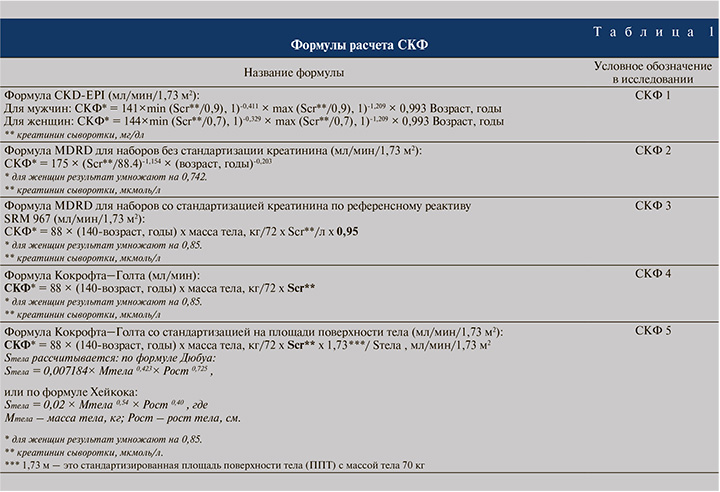
- расчет скорости клубочковой фильтрации (СКФ) по формулам (табл. 1);
- КТ мочевыводящих путей с контрастированием с определением максимального размера кисты;
- вычислялись объем паренхимы почки и объем потерянной (атрофированной) паренхимы с оценкой компрессии и деформации кистой чашечно-лоханочной системы (ЧЛС);
- динамическая нефросцинтиграфия с расчетом суммарной СКФ и СКФ каждой почки в отдельности;
- за объем атрофированной паренхимы почки принимались объем кратера кисты почки при экзофитном росте кисты и объем кисты при ее интрапаренхиматозном расположении, фактический объем почечной паренхимы рассчитывался методом сегментации (рис. 1а, 1б). Сначала определялась площадь почечной паренхимы путем контурирования последней на аксиальном срезе, после чего объем среза автоматически рассчитывался программой путем умножения площади паренхимы на длину среза. Объем почечной паренхимы рассчитывался путем суммирования объемов всех сегментов (срезов). Объем кратера рассчитывался также методом сегментации. При контурировании кратера на аксиальном срезе внешний контур кратера формировался путем дорисовывания исследователем кривой линии, продолжающей внешний контур почки (рис. 1а, 1б). Исходный объем почечной паренхимы рассчитывался путем суммирования фактического объема почечной паренхимы и объема «кратера» кисты почки.


Критерии включения:
- взрослые пациенты с простой кистой почки Босниак I–II;
- солитарная киста почки;
- одностороннее поражение.
Критерии невключения:
- невозможность выполнения КТ с контрастированием;
- кисты Босниак IIF, III, IV;
- признаки хронической болезни почек (ХБП) 3a и выше;
- билатеральные кисты;
- множественные кисты почки;
- синусные кисты почки;
- кисты максимальным размером менее 2 см;
- единственная почка;
- гломерулонефрит;
- амилоидоз;
- сахарный диабет;
- аутоиммунные заболевания;
- мочекаменная болезнь;
- опущение или дистопия почки;
- вторично сморщенная или нефункционирующая почка;
- гипоплазия почки;
- добавочная почка;
- операции на почках и верхних мочевыводящих путях;
- аномалии развития почек и верхних мочевыводящих путей с нарушением оттока мочи;
- кальциноз почечных артерий;
- стеноз почечных артерий;
- гипертоническая нефропатия;
- тяжелые сопутствующие заболевания, требующие проведения:
- химиотерапии;
- длительной (более 10 дней) терапии антибиотиками за последний месяц;
- длительного (более 7 дней) приема НПВС за последний месяц;
- диагностические и лечебные манипуляции, сопровождающиеся введением рентген-контрастных препаратов в течение 30 дней.
После получения результатов проводился анализ симметричности функции обеих почек путем сравнения СКФ пораженной и здоровой почек, анализировалась связь между наличием простой кисты почки и снижением ее функции по сравнению со здоровой почкой, а также связь между максимальным размером кисты почки и снижением ее функции по сравнению со здоровой почкой. Также анализу подвергалась связь между объемом атрофированной паренхимы почки и снижением ее функции по сравнению со здоровой почкой. Наряду с этим проводился анализ соответствия показателей суммарной СКФ, полученных при сцинтиграфии, и показателей СКФ, рассчитанных при помощи формул, указанных в табл. 1.
Статистические методы
При анализе количественных данных проведено предварительное тестирование переменных на нормальность распределения с помощью теста Шапиро–Уилка. В случае нормального распределения параметр представлялся в виде арифметического среднего (M) + стандартного отклонения (SD) (mean±std), при отклонении от нормального распределения параметр представлялся в виде медианы (Me) и 1-го и 3-го квартилей (Q1, Q3)) (median (q1; q3)). При необходимости для оцениваемых параметров строились 95-процентные доверительные интервалы, которые приводятся в виде [нижняя граница ДИ; верхняя граница ДИ].
При сравнении параметров в случае нормального распределения использовался парный t-тест Стьюдента, в случае логарифмического нормального распределения t-тест применялся к логарифмическому преобразованию исходного параметра. При иных распределениях применялся W-критерий Вилкоксона для связных выборок. Значения р округлялись до трех десятичных знаков. В случае p-значений с первым ненулевым знаком после запятой, не попадающим в указанную точность, p-значение приводилось в виде ноля. Для анализа параметров, влияющих на СКФ пораженной почки, использована обобщенная линейная модель (GLM). Для анализа вероятности значимого снижения СКФ пораженной почки, а также для факторов, влияющих на эту вероятность, построена модель многофакторной логистической регрессии. Для оценки качества модели использован ROC (receiver operating characteristic)-анализ.
Результаты исследования. Для анализа были доступны данные 109 пациентов, из которых 62 (56,9%) пациента были мужского пола и 47 (43,1%) – женского. Их средний возраст составил 62 (54;68) года, индекс массы тела (ИМТ) – 28,16±4,07 кг/м², показатель креатинина крови – 87,4±20,93 мкмоль/л. У 55 (50,5%) пациентов киста почки располагалась слева, у 54 (49,5%) – справа. У 43 (39,5%) пациентов киста локализовалась в верхнем сегменте почки, у 42 (38,5%) – в среднем и у 24 (22%) пациентов – в нижнем сегменте почки. У 53 (48,6%) определялась деформация и сдавление ЧЛС почки кистой.
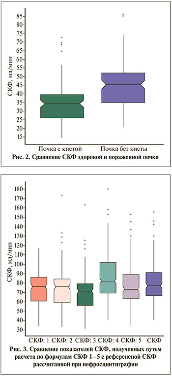
Максимальный размер кисты составил 80 (66; 97) мм. При этом наибольший максимальный размер кисты был равен 201 мм, минимальный – 46. Объем паренхимы пораженной почки составил 174 (137;206) мл, объем атрофированной или утраченной паренхимы (объем кратера кисты) – 49 (27;71) мл, а доля утраченной паренхимы – 28% (19%; 37%). Суммарная СКФ составила 77,07 (66,8; 90,8) мл/мин. СКФ здоровой почки составила 45,49 (35,03;52,07) мл/мин, а СКФ пораженной кистой почки – 34,46 (25,97; 39,63) мл/мин. Средняя разница СКФ здоровой и пораженной кист почки составила 11 [8,70; 13,44] мл/мин и была статистически значимой (р=0) (рис. 2).

Сравнение показателей СКФ, полученных по формулам 1–5, с референcными значениями суммарной СКФ, полученных при сцинтиграфии, показал что формулы СКФ1, СКФ3 и СКФ4 дают статистически значимое (на уровне 5%) систематическое отклонение от референсных значений СКФ. Формула СКФ2 дает статистически значимое (на уровне 10%) систематическое отклонение от референсной СКФ. Таким образом, формулу СКФ5 можно определить как формулу, дающую значения СКФ, наиболее приближенные к референсным (рис. 3).
Корреляционный анализ показал, что доля утраченной паренхимы статистически значимо коррелировала с максимальным размером кисты: ρ=0,37 с 95% ДИ [0,20; 0,52] (р=0). Поэтому в дальнейших моделях рассматривался только показатель доли утраченной паренхимы, а максимальный размер кисты был исключен из использования в моделях.
В качестве обобщенной линейной модели для логарифма СКФ пораженной почки выбрана модель со следующими параметрами: пол, возраст, ИМТ, логарифм доли утраченной паренхимы и наличие компрессии или деформации ЧЛС почки кистой. Статистический анализ показал, что значимым (на уровне значимости 5%) параметром, отрицательно влияющим на изменение СКФ почки с кистой, является доля утраченной паренхимы (табл. 2).

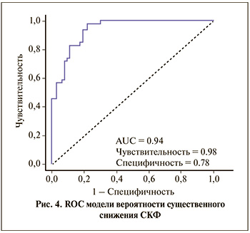
Для оценки вероятности существенного снижения СКФ (более чем на 10 мл/мин) и выявления факторов, статистически значимо влияющих на эту вероятность, построена модель многофакторной логистической регрессии. По результатам анализа, статистически значимым фактором, влияющим на вероятность существенного снижения СКФ, является доля утраченной паренхимы почки. Увеличение доли утраченной паренхимы на 1% ведет к увеличению шанса существенного снижения СКФ в 1,13 раза. То есть увеличение данного показателя на 10% увеличивает вероятность снижения СКФ пораженной почки на 10 мл/мин в 3,39 раза, а увеличение потери паренхимы на 20% увеличивает вероятность снижения СКФ пораженной почки на 10 мл/мин в 11,52 раза (табл. 3).

Качество выбранных параметров модели анализировалось расчетом AUC (area under curve – площади под кривой) для ROC. На рис. 4 приведена ROC для модели вероятности успешного лечения. AUC для такой ROC равна 0,94, что говорит об отличном качестве модели.

Обсуждение. Полученные нами данные поднимают вопрос о целесообразности выполнения динамической нефросцинтиграфии с целью оценки снижения функции пораженной почки для определения показаний к хирургическому лечению кисты почки. Снижение функции почки в результате сдавления и атрофии паренхимы было показано в экспериментальной работе Gomez и соавт., которые по результатам гистологического исследования препаратов почек с кистами продемонстрировали признаки атрофии почечной паренхимы с резким уменьшением количества нефронов в зоне сдавления паренхимы почки кистой [17]. Al-Said и соавт. в исследовании, охватившем 561 пациента с кистами почек, отметили снижение функции почек даже при наличии единичных кист [19]. В большинстве работ на эту тему изучена связь снижения функции почки с размером кисты. Так, Kwon и соавт. продемонстрировали снижение функции почки у 31 (60,2%) из 50 пациентов при среднем размере кисты 7,2 см [18]. J. Chen и соавт. в крупном когортном исследовании, в котором представлены результаты наблюдения за 4274 пациентами с кистами почек в течение 5 лет, отметили, что размер кисты более 2,2 см сопряжен с рисками более стремительного снижения функции почек [16]. Исходя из представления об относительной симметрии функции почки здорового человека, согласно которой каждая почка обладает одинаковыми показателями СКФ, результаты нашего исследования подтвердили данные мировой литературы, показав, что рост кисты почки сопровождается снижением СКФ пораженной почки. Наряду с этим в нашем исследовании было показано, что рост кисты приводит к атрофии и утрате почечной паренхимы, что является наиболее вероятным механизмом снижения функции почки. Этот тезис косвенно подтверждается результатами работы Wu и соавт., показавшими, что снижение почечной функции наблюдалось в 10 раз чаще в группе пациентов с прогрессивно растущими кистами по сравнению с группой пациентов со «стабильными» кистами (23,3 против 2,4%) [13]. В среднем в выборке пациентов, представленной в нашем исследовании, рост кисты привел к утрате 49 мл (28%) паренхимы почки, что привело к снижению СКФ на 11 мл/мин по сравнению со здоровой почкой. Статистический анализ также выявил прямую связь между максимальным размером кисты, объемом утраченной паренхимы и СКФ пораженной почки. Таким образом, согласно данным нашего исследования, рост кисты почки может приводить к снижению СКФ пораженной почки, что может рассматриваться в качестве относительного показания к хирургическому лечению простой кисты почки. По этой причине пациентам с простой кистой почки на основании полученных результатов можно рекомендовать выполнять динамическую нефросцинтиграфию с целью оценки снижения функции пораженной почки для определения показаний к хирургическому лечению кисты почки. В качестве показаний к выполнению динамической нефросцинтиграфии мы рекомендуем использовать показатель доли утраченной паренхимы в 20% и более (рассчитанной методом сегментации), поскольку потеря одной пятой объема почечной паренхимы увеличивает вероятность существенного снижения СКФ более чем в 10 раз.
Ограничения исследования
Ограничением нашего исследования можно считать сниженные показатели медианы суммарного СКФ исследуемых, которая, несмотря на строгие критерии исключения из исследования, отсеивавших пациентов с самыми распространенными причинами снижения почечной функции, может быть связана с другими причинами нарушения почечной функции. Поскольку в клинической практике не редки случаи множественных кист почек и их двустороннего расположения, другим ограничением могут считаться критерии включения в исследование, включавшие исключительно пациентов с солитарной кистой почки и односторонним характером поражения. В КДЦ ГКБ им. С. И. Спасокукоцкого обращаются пациенты, направляемые из поликлиник, чаще всего с целью решения вопроса о хирургическом лечении кисты почки. Данное обстоятельство объясняет включение в исследование преимущественно пациентов с крупными размерами кисты почки. Существуют данные, что объем правой и левой почек у здоровых людей могут различаться в среднем на 5 мл [22]. Несмотря на столь незначительное различие, подобная анатомическая асимметрия может приводить к разнице СКФ правой и левой почек в 1–2 мл/мин, что также не учитывалось в нашем исследовании.
Выводы. Результаты нашего исследования показали, что рост кисты почки вызывает атрофию почечной паренхимы и снижение СКФ пораженной почки. Увеличение объема атрофированной паренхимы приводит к снижению СКФ пораженной почки. Согласно полученным результатам, утрата 20% почечной паренхимы может рассматриваться в качестве показания к выполнению динамической нефро-сцинтиграфии. Формула Кокрофта–Голта со стандартизацией на площади поверхности тела позволяет рассчитывать показатели СКФ, наиболее приближенные к значениям СКФ, полученные при сцинтиграфии и, следовательно, может быть рекомендована в качестве оптимальной формулы для расчета СКФ в ежедневной клинической практике.


