
Реномедуллярные интерстициальные опухоли (РИО), или медуллярные фибромы, – доброкачественные мезенхимальные новообразования у взрослых, нередко случайно выявляемые при проведении аутопсий [1, 2]. Так, по данным M. R. Martin и A. J. Tiltman (1976), на секционном материале данные опухоли встречались в 36% случаев у лиц старшего и пожилого возраста [3]. Другие авторы указывают на крайне редкую частоту РИО и приводят описание 41 наблюдения данных опухолей [4]. В целом в отечественной и зарубежной литературе отсутствуют сводные данные, посвященные частоте встречаемости РИО.
Возникновение и развитие РИО представляют особый интерес. Так, по мнению некоторых авторов, данные опухоли возникают из реномедуллярных интерстициальных клеток, в роли которых могут выступать фибробласты и миофибробласты [1, 2]. Известно, что данные клеточные популяции участвуют в процессах синтеза и секреции коллагеновых волокон протеогликанов – основных компонентов соединительной ткани. Кроме того, интерстициальные клетки мозгового слоя почки могут принимать участие в регуляции уровня ренина и бикарбонатов, следовательно, влиять на системное артериальное давление. В связи c этим отдельные работы, посвященные изучению морфологических и иммуногистохимических особенностей интерстициальных клеток почек, заслуживают пристального внимания [4, 5].
Патогистологическое исследование РИО демонстрирует вариабельное строение. Некоторые авторы выделяют два гистологических варианта РИО: клеточный и фиброзный [2, 3].
Клеточный вариант РИО представлен преобладанием пролиферирующих интерстициальных клеток – фибробластов в мозговом слое почки с их локализацией между медуллярными канальцевыми структурами (дистальными канальцами, собирательными трубками).
Фиброзный вариант РИО содержит массу фибробластов, миофибробластов, расположенных среди фиброзной отечной интерстициальной ткани, окружающей канальцевые структуры. Строма опухоли может содержать белковые эозинофильные массы, толстые коллагеновые волокна III типа.
Работами отдельных авторов показано, что интерстициальные клетки РИО могут содержать рецепторы гормонов к эстрогену, прогестерону. По-видимому, это может свидетельствовать о роли данных клеток в обмене гормонов, однако это требует дальнейшего молекулярно-биологического исследования [4].
В связи с редкостью РИО в клинической и патологоанатомической практике приводим собственное секционное наблюдение.
Пациент 49 лет. Из истории болезни известно, что в течение 12 лет больной страдал сахарным диабетом 2-го типа стадии компенсации, артериальной гипертензией II стадии с периодическими подъемами артериального давления до 160/100 мм рт.ст. В связи с данными заболеваниями больной в течение ряда лет получал корригирующую терапию манинилом (2,5 мг/сут), эналаприлом (2,5 мг/сут). На фоне гипертонического криза у больного возник ишемический инсульт серого и белого веществ правого полушария головного мозга с последующим развитием, прогрессированием отека и набухания, что послужило непосредственной причиной смерти.
На вскрытии были обнаружены признаки ишемического размягчения серого и белого веществ головного правой теменно-височной области, признаки гипертонической болезни: гипертрофия миокарда левого желудочка сердца (масса сердца – 430 г, толщина стенки левого желудочка – 3,4 см), микроангиопатия сосудов почек в виде утолщения, склероза, гиалиноза стенок приносящих и выносящих артериол, капиллярных петель клубочков.
В мозговом слое правой почки на вершине пирамидки имелся опухолевый белый узелок диаметром 0,4 см плотной консистенции с четкими границами с окружающей мозговой тканью почки.
При гистологическом исследовании почки использовали окраски гематоксилином и эозином, пикро-Маллори для выявления коллагеновых и эластических волокон, по Хочкиссу (ШИК-реакция) для обнаружения гликозаминогликанов, метенамином серебра для оценки базальных мембран капилляров клубочков, артерий, артериол, стромы опухоли.
Капиллярные петли клубочков почек были утолщены за счет ШИК-позитивных веществ, имели «лапчатый вид», имелись признаки склероза и гиалиноза. Стенки приносящей и выносящей артериол были неравномерно утолщены за счет масс гиалина, просветы сосудов сужены (рис. 1). Эти изменения указывали на наличие микроангиопатии, гломерулопатии, вероятно обусловленные имевшимися заболеваниями.
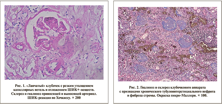
Местами определялись тотальный склероз и гиалиноз клубочков с признаками хронического тубулоинтерстициального нефрита с резкой атрофией канальцевого аппарата в строме – признаки микроангиопатии и очагового нефросклероза (рис. 2).
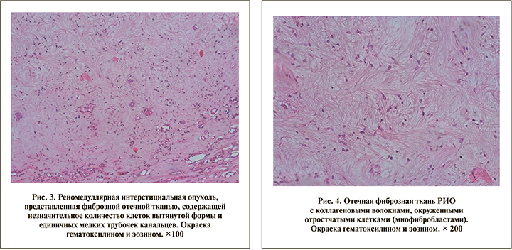
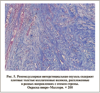
Реномедуллярная интерстициальная опухоль была представлена узлом, содержащим фиброзную ткань с признаками отека, в которой встречались отдельные клетки вытянутой формы. В периферических отделах опухоли имелись единичные атрофичные канальцевые структуры (рис. 3). При большом увеличении ткань опухоли была представлена переплетающимися между собой тонкими и толстыми коллагеновыми волокнами, окруженными отростчатыми клетками – миофибробластами (рис. 4, 5).
 Таким образом, приведенное секционное наблюдение пациента 49 лет демонстрирует редкую опухоль почки, имеющей строение мезенхимальной реномедуллярной интерстициальной опухоли (РИО) со своими особенностями. Среди них – преобладание в опухоли клеточного компонента, представленного отростчатыми элементами (фибробласты, миофибробласты), окруженными коллагеновыми волокнами интерстиция.
По-видимому, данные клетки могут принимать участие в регуляции уровня ренина и бикарбонатов, обмена компонентов соединительной ткани (коллагеновые волокна, протеогликаны), гормонов (эстрогена, прогестерона). Полагая, что данные клетки опухоли регулируют синтез и секрецию представленных веществ, можно думать о многочисленных клинических проявлениях РИО. Вероятнее всего, к таковым можно отнести повышение артериального давления, водно-электролитный дисбаланс, дисгормональные расстройства в связи с наличием рецепторов к эстрогену, прогестерону в ткани опухоли, метаболические расстройства (сахарный диабет). Однако эти положения требуют дальнейшего клинико-морфологического и иммуногистохимического исследований.