
Введение. Изменения, возникающие в нормальных тканях после проведения лучевой терапии по поводу злокачественных новообразований, несмотря на развитие методов конформного облучения, до сих пор остаются нерешенной проблемой современной радиационной онкологии [1].
При облучении органов малого таза по поводу опухолей женской репродуктивной системы органами, подверженными высокому риску нежелательных эффектов лучевой терапии, являются мочевой пузырь и прямая кишка. Для практической урологии наиболее важное клиническое значение имеют поздние лучевые повреждения мочевого пузыря, которые появляются спустя 3 и более месяцев после окончания облучения, а иногда через несколько лет после лучевой терапии. Выраженность их может варьироваться от относительно незначительных функциональных нарушений до тяжелых осложнений, оказывающих влияние на качество жизни пациентов, вплоть до инвалидизации [2].
Трудность ведения таких пациентов для практикующего врача-уролога известна. Один из путей решения проблемы – развитие системы профилактики тяжелых лучевых повреждений и ее своевременное проведение, другой – оптимизация лечения возникших осложнений.
Оценить состояние соединительнотканного матрикса мочевого пузыря до облучения и, соответственно, выявить группу риска развития тяжелых лучевых повреждений до настоящего времени не представлялось возможным.
Традиционное использование стандартных методов гистологического исследования исключает возможность прижизненной оценки органа и не позволяет получить диагностически значимую информацию о состоянии ведущего компонента тканевого метаболизма в стенке мочевого пузыря – соединительнотканного матрикса. Современные оптические методы визуализации структуры коллагена и эластина [3] позволили вернуться к прицельному исследованию лучевых повреждений проблемных органов малого таза в целом и мочевому пузырю в частности на новом уровне.
Цель исследования: определить роль структурных изменений внеклеточного матрикса мочевого пузыря в возникновении побочных эффектов лучевой терапии разной степени тяжести.
Материалы и методы. Анализ состояния соединительнотканного матрикса выполнен по 126 изображениям, полученным с гистологических срезов биоптатов мочевого пузыря 12 пациентов, классифицированных согласно клинической картине заболевания и степени тяжести побочных эффектов облучения (по шкале оценки поздних лучевых повреждений RTOG/EORTC [4]): при II степени тяжести проанализировано 36 изображений (4 пациента), при третьей – 50 (n=5), при четвертой – 40 (n=3). Контрольные изображения нормального мочевого пузыря (n=23) получены из секционного материала трех образцов ткани нормального мочевого пузыря, взятых post mortem.
Для изучения соединительнотканного матрикса при осложнениях разной степени тяжести лучевого поражения мочевого пузыря применен метод нелинейной микроскопии в режимах регистрации сигнала генерации второй гармоники (ГВГ) и двухфотонного возбуждения автофлуоресценции (ДВАФ). Использование метода нелинейной микроскопии позволило получить информацию о состоянии и «упаковке» пучков коллагеновых и эластических волокон без окрашивания гистологического препарата.
Исследование неокрашенных депарафинированных гистологических срезов методом нелинейной микроскопии проводилось на инвертированном лазерном сканирующем микроскопе LSM 510 («Carl Zeiss», Германия). В качестве источника возбуждающего излучения был использован короткоимпульсный фемтосекундный лазер MAI TAI HP («Spectra Physics», США) с частотой следования импульсов 80 МГц и длительностью порядка 100 фс.
Возбуждение осуществлено на длине волны 800 нм, регистрация сигнала ГВГ от коллагеновых структур проведена в диапазоне 362–415 нм (изображение в зеленом цвете), сигнала ДВАФ от эластических волокон – в диапазоне 512–576 нм. Изображения размером 318×318 мкм (1024×1024 пиксела) строились с помощью масляно-иммерсионного объектива Plan-Neofluar 40x/1.3 («Carl Zeiss», Германия).
Для верификации полученных изображений исследованы гистологические срезы фрагментов мочевого пузыря, сделанные из тех же парафиновых блоков, что и для нелинейной микроскопии, окрашенные гематоксилином и эозином.
Результаты
Клинические проявления лучевого поражения мочевого пузыря. При II степени тяжести пациенты жаловались на умеренно учащенное мочеиспускание; при цистоскопии выявлены единичные телеангиэктазии преимущественно на задней стенке при нормальной емкости мочевого пузыря. При III и IV степенях тяжести лучевого поражения значительное учащение мочеиспускания сопровождалось резями, болью. Больные жаловались на наличие крови в моче. При цистоскопии фиксировались снижение емкости мочевого пузыря, наличие петехиальных кровоизлияний, преимущественно на задней стенке. При IV степени выявлены язвы с фибринозным налетом, некрозами, инкрустацией солями.
Гистологическое исследование. При лучевом цистите вне зависимости от степени тяжести во внутренних слоях стенки (слизистой оболочке, подслизистом слое) было обнаружено диффузное неравномерное разрастание фиброзной ткани. Иногда выявляли слабовыраженную воспалительную инфильтрацию из лимфоцитов, плазматических клеток, макрофагов, иногда зоны отека и полнокровия. Покровный уротелий с участками атрофии, местами очаговой гиперплазии.
Нелинейная микроскопия. Результаты исследования гистологических срезов мочевого пузыря методом нелинейной микроскопии показали, что структурные изменения соединительнотканного матрикса при разной степени тяжести лучевых осложнений различаются.
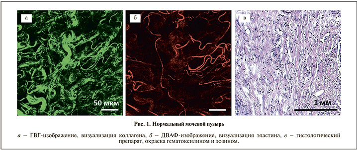
Коллагеновые волокна в нормальном мочевом пузыре имеют извитую форму, местами собраны в пучки (рис. 1, а). Коллагеновые волокна и пучки расположены рыхло и сопровождаются эластическими волокнами (рис. 1, б).
При II степени тяжести осложнений лучевой терапии структура коллагеновых волокон сохранялась на фоне их уплотнения (рис. 2, а1). При III степени тяжести наблюдали их выраженную дезорганизацию, размытость, волокна были сильно извитыми, не имели выделенного направления (рис. 2, б1).
Эластические волокна в норме визуализируются как тонкие сильно извитые волокна одинакового диаметра [5]. Волокна четко очерчены, не имеют преобладающего направления в пространстве (рис. 1, б). При II и III степенях тяжести осложнений лучевой терапии наблюдалась потеря характерной для них в норме извитой формы укладки, мозаично встречались сильно разветвленные участки (рис. 2, а2, б2).
При III и IV степенях тяжести лучевых поражений выявлены очаги бесструктурной ткани с высоким сигналом ГВГ (рис. 2, б1; рис. 3, б1, области, обозначенные красными пунктирными кругами), что свидетельствует о высокой степени анизотропии и плотности данных образований, но без признаков организации. Такая особенность сигнала ГВГ может соответствовать отложению масс гиалина по ходу коллагеновых волокон, поскольку известно, что в процессе дезорганизации соединительной ткани на фоне какого-либо поражения происходит деполимеризация гликозаминогликанов, разрушение коллагеновых волокон и пропитывание их плазменными белками, в результате коллаген образует с фибриногеном и другими веществами плотные нерастворимые соединения [6].
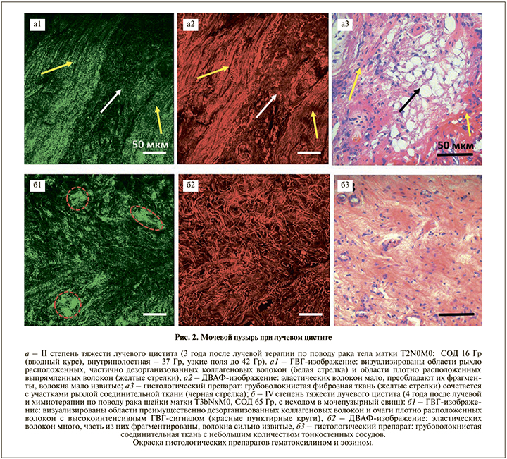
При сопоставлении описанных участков с гистологическими препаратами выявлено, что при окраске гематоксилином и эозином на фоне фиброзной ткани они четко не дифференцируются (рис. 2, б3; рис. 3, б3).
Таким образом, в целом получена корреляция клинического проявления лучевого поражения мочевого пузыря по степени тяжести с данными нелинейной микроскопии. Можно сделать вывод, согласно которому клинические проявления различных по степени тяжести побочных эффектов лучевой терапии при радиационном поражении мочевого пузыря зависят от состояния внеклеточного матрикса.
«Горячими зонами», максимально страдающими от облучения, являются задняя стенка, мочепузырный треугольник и шейка мочевого пузыря, в то время как внеклеточный матрикс боковых и передней стенок изменяется в меньшей степени. В ранее проведенных исследованиях ткани мочевого пузыря, взятых из разных зон, выявлено, что степень поражения ткани на фоне лучевой терапии разная, даже в максимально страдающих зонах носит характер мозаичности [5, 7]. То есть патогенетически оправдано проведение различных курсов комплексного лечения с целью сохранения функциональных резервов мочевого пузыря, предотвращения прогрессирования снижения его емкости даже при тяжелых степенях осложнений лучевой терапии.
Ниже на клиническом примере пациентки, имевшей осложнения со стороны мочевого пузыря после лучевой терапии III степени тяжести, продемонстрирован разный уровень сохранности ткани мочевого пузыря.
П а ц и е н т к а С. 79 лет. В 2010 г. пациентке выполнена экстирпация матки с придатками по поводу рака шейки матки Т1bN0M0. В 2011 г. проведен курс лучевой терапии (дистанционная лучевая терапия СОД 40 Гр, РОД 2 Гр, внутриполостная лучевая терапия СОД 25 Гр, РОД 5 Гр). С 2012 г. появились неудержимые позывы на мочеиспускание, рези при мочеиспускании, боли над лоном, диарея. При цистоскопии в 2014 г. выявлен язвенный цистит на фоне лучевого поражения задней стенки мочевого пузыря. Нерегулярная консервативная терапия приносила временный эффект, хотя язва зажила. В 2016 г. при очередном обращении пациентки за медицинской помощью по данным УЗИ выявлено уменьшение объема мочевого пузыря до 70 см3.
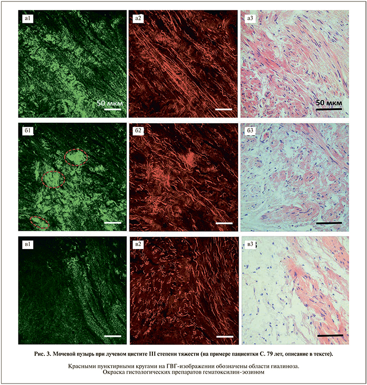
При цистоскопии, выполненной в связи с подозрением на развитие вторичного инфильтративного рака мочевого пузыря 27.01.2016, проведена трансуретральная резекция в зоне мочепузырного треугольника и мультифокальная биопсия из стенок. Наряду со стандартным морфологическим исследованием гистологические препараты изучены методом нелинейной микроскопии (рис. 3). Выявлена разная степень сохранности внеклеточного матрикса стенок мочевого пузыря. На ГВГ-изображениях из боковых стенок коллаген спиралевидный, структура большинства волокон сохранена (рис. 3, а1), на ДВАФ-изображениях эластических волокон много, часть из них имеют извитой ход, часть прямые (рис. 3, а2), что свидетельствует в целом о хорошей упругости субэпителиальной ткани мочевого пузыря в данной зоне.
На ГВГ-изображении из задней стенки мочевого пузыря пучки коллагеновых волокон несколько размытые (рис. 3, б1), присутствуют гомогенные, ярко окрашенные участки – показатель наличия анизотропной среды. Вероятно, это очаги фибриноидного некроза с исходом в гиалиноз (рис. 3, б1). На ДВАФ-изображении видно, что эластические волокна тонкие, имеют направление, аналогичное таковому коллагеновых волокон (рис. 3, б2). Такое состояние волокон лежит в основе дисфункциональности –недостаточной упругости субэпителиальной ткани мочевого пузыря в данной зоне и является причиной клинических проявлений: снижения емкости, болей, дизурии и пр.
ГВГ- и ДВАФ-изображения пограничной зоны между боковой и задней стенками мочевого пузыря представлены на рис. 3, в1 и в2 соответственно. На ГВГ-изображении видна граница между мышечными и коллагеновыми волокнами, последние несколько размытые, но ровные и прямые, организованные, в основном плотно уложенные, с небольшими включениями более толстых и извитых пучков (рис. 3, в1). Эластические волокна тонкие, чуть извитые, имеют направление, аналогичное коллагеновым (рис. 3, в2). Все волокна в поле зрения имеют однонаправленное положение. Гистологически соединительная ткань рыхлая, коллагеновые волокна тонкие, их структура в целом сохранена. Справа на изображении определяется небольшое количество атрофированных гладкомышечных волокон (рис. 3, в3).
Обсуждение. Основным направлением профилактики тяжелых осложнений лучевой терапии в настоящее время является использование высокотехнологичных конформных методик облучения, которые позволяют создать высокий градиент дозы на границе мишени и включить в объем облучения минимальный объем нормальных тканей. Однако внедрение таких подходов не всегда позволяет избежать высоких радиационных нагрузок на органы, расположенные вблизи патологического очага. При облучении новообразований органов малого таза (рак тела матки, рак шейки матки, рак предстательной железы, рак прямой кишки) основным органом риска является мочевой пузырь, доза облучения на отдельные зоны которого при проведении сочетанной лучевой терапии может существенно превышать толерантные.
Деструкция коллагеновых волокон любой природы играет значимую роль в патогенезе повреждения мочевого пузыря и зачастую является причиной последующих функциональных расстройств. С ней связаны изменения сосудистой стенки и последующая облитерация сосудов, которая приводит к гипоксии и дистрофическим изменениям тканей. Такие изменения показаны нами ранее при исследовании мочевого пузыря методами лазерной доплеровской флоуметрии [8] и кросс-поляризационной оптической когерентной томографии [9]. Известно, что повышенный риск развития тяжелых радиационно-индуцированных повреждений органов малого таза имеют пациенты с хроническими воспалительными заболеваниями мочевого пузыря [10]. Таким образом, исходное состояние соединительнотканного матрикса мочевого пузыря до лучевой терапии, а также структурные изменения, возникшие в процессе развития осложнений лучевой терапии, важны для сохранения его функциональных резервов.
Метод нелинейной микроскопии с верифицирующим гистологическим исследованием позволил выявить особенности пространственной и структурной организации внеклеточного матрикса мочевого пузыря при разной степени лучевого поражения. Полученные в нашем исследовании клинические результаты (нарушение структуры волокон и снижение интенсивности сигнала генерации второй гармоники) соответствуют изменениям, показанным ранее в экспериментальных исследованиях [5, 11]. В работе на примере исследования мочевого пузыря при осложнениях лучевой терапии III степени тяжести показан разный уровень сохранности его соединительнотканного матрикса, что свидетельствует о необходимости проведения длительной комплексной патогенетической терапии, направленной на улучшение трофических процессов в стенке органа. Важность знания исходного состояния соединительнотканного матрикса мочевого пузыря для лечащего врача состоит в том, что оно может стать основой прогнозирования степени тяжести лучевых осложнений.
В основу профилактики лучевых осложнений до начала лучевой терапии должна быть включена обязательная консультация врачом-урологом, преследующая цель выявления и лечения хронических воспалительных заболеваний мочевого пузыря. Своевременное лечение позволит уменьшить дистрофические процессы в стенке мочевого пузыря при облучении, что в свою очередь будет влиять на сохранность его функциональных резервов. Другой путь – оптимизация лечения возникших осложнений.
Заключение. Исследование образцов тканей мочевого пузыря, взятых из разных зон, проведенное методом нелинейной микроскопии в режимах ГВГ и ДВАФ, выявило, что степень структурных изменений соединительнотканного матрикса в послелучевой период разная и коррелирует со степенью тяжести лучевых осложнений, диагностированных клинически. Результаты данного исследования могут быть основополагающими в системе профилактики тяжелых радиационных повреждений мочевого пузыря при лучевой терапии онкологической патологии органов малого таза.
Финансирование исследования
Работа выполнена при финансовой поддержке гранта РФФИ № 16-07-00655 и гранта РФФИ № 16-02-00670.