Введение. Мочекаменная болезнь (МКБ) является распространенным и наиболее дорогостоящим из урологических заболеваний во всех регионах планеты. ССЫЛКУ Изучение распространенности видов мочевых камней на различных территориях страны и мира играет большое значение в предсказании нагрузки на системы здравоохранения в целом и урологическое сообщество в частности, в том числе и в плане расчетов вероятности рецидивирования заболевания даже на фоне эффективно проводимой метафилактической терапии.
Цель: в связи с вышеуказанным нами предпринята попытка оценки распространенности различных видов мочевых камней в различных регионах Российской Федерации, Белоруссии, Казахстана и особенности изменений состава мочевых камней в зависимости от возраста и пола.
Материалы и методы. Исследование основано на данных изучения химического состава 6787 мочевых камней, представленных компанией «ИНВИТРО» в обезличенном виде за 2018–2021 гг. Исследование химического состава камней проводилось методами инфракрасной спектроскопии и/или рентгеновской дифракции. Все полученные данные были обработаны в программе Excel, 2007. Результаты качественных признаков выражены в абсолютных числах с указанием долей (%). Сравнение номинальных данных в группах проводилось при помощи критерия χ2 Пирсона.
В тех случаях, когда число ожидаемых наблюдений в любой из ячеек четырехпольной таблицы было менее 10, для оценки уровня значимости различий использовался точный критерий Фишера. Статистически значимыми считались различия при p≤0,05.
Результаты исследования. Из 6787 исследованных камней однокомпонентные по минеральному составу камни выявлены у 448 человек обоего пола, что составило 6,6%, двухкомпонентные – в 4005 (59%) наблюдениях, трех- и более компонентные (многокомпонентные) – в 2334 (34,4%). Из общего числа исследованных камней 538 были получены у детей и подростков в возрасте до 25 лет. В этой группе однокомпонентные камни выявлены в 28 случаях, что составило 5,2 %, двухкомпонентные – в 298 (55,4 %), трех- и более компонентные камни обнаружены в 212 (39,4%) случаях. Таким образом, распределение камней по указанным категориям среди взрослых и детей было примерно одинаковым.
Однако среди однокомпонентных камней отмечена существенная разница в минеральном составе у детей и взрослых. В обеих группах преобладали кальцийсодержащие однокомпонентные камни – у 73,4% взрослых и у 71,4% детей. Наиболее распространенным минералом являлся кальция оксалат моногидрат (СОМ) – 65,8% у взрослых и 46,4% у детей. У взрослых из кальцийнесодержащих камней преобладали камни из мочевой кислоты и дигидрата мочевой кислоты (МК/ДМК) – 18,8% и достоверно преобладали среди мужчин, среди детей – «инфекционные» камни – в 25%.
Обращает на себя внимание высокая частота обнаружения инфекционных камней из струвита и урата аммония у девочек (100%).
При оценке распределения по регионам среди взрослого населения повсеместно преобладали камни из кальция оксалата моногидрата (СОМ), камни из карбонатапатита (СаР) чаще встречались у пациентов в Южном, Уральском и Приволжском федаральных округах (ФО), камни из мочевой кислоты (МК/ДМК) – в республиках Крым, Казахстан; Северо-Кавказском, Дальневосточном ФО и в Москве, урат аммония – у больных в Республике Белоруссия и Приволжском ФО, струвит – в республиках Казахстан, Белоруссия; Сибирском, Приволжском и Северо-Западном ФО и Москве. По детскому контингенту такой информации нам получить не удалось.
При оценке распределения камней по гендерному признаку отмечено, что камни из СОМ и камни из МК/ДМК среди мужчин встречались чаще, чем среди женщин, – 64,4 против 35,6% (р<0,001) и 65 против 35% (р<0,001) соответственно. Камни из струвита чаще выявляли у женщин (80%), чем у мужчин (p<0,001), камни из СаР незначительно чаще встречались у мужчин – 47 против 53% у женщин (р=0,397) (рис. 1).

У детей камни из кальция оксалата моно- и дигидрата (СОМ и СОD) встречались примерно в одинаковом соотношении между мальчиками и девочками, камни из СаР – чаще у девочек (<0,001), из мочевой кислоты (МК) – только у мальчиков.
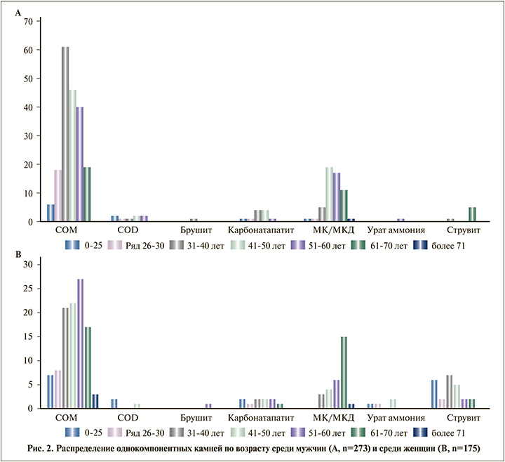
При оценке соотношения типов мочевых камней с возрастом отмечена высокая распространенность СОМ в возрастной период от 26 до 70 лет среди мужчин и женщин. Камни из МК наиболее часто встречались среди мужчин в возрастной период от 31 года до 70 лет, среди женщин – от 41 года до 70 лет. Струвитные камни в женской популяции наиболее часто выявлялись в детском возрасте и в возрасте 31–50 лет (период сексуальной активности). Отмечено увеличение струвитного камнеобразования среди мужчин старше 61 года, возможно, обусловленное развитием осложнений, связанных с доброкачественной гиперплазией предстательной железы (рис. 2).

Двух- и более компонентные камни представляли собой различные композиции минералов с преобладанием кальция и встречались как среди взрослых, так и среди детей. При оценке конкрементов данных групп на первое место ставился минерал с наибольшим присутствием в составе камня.
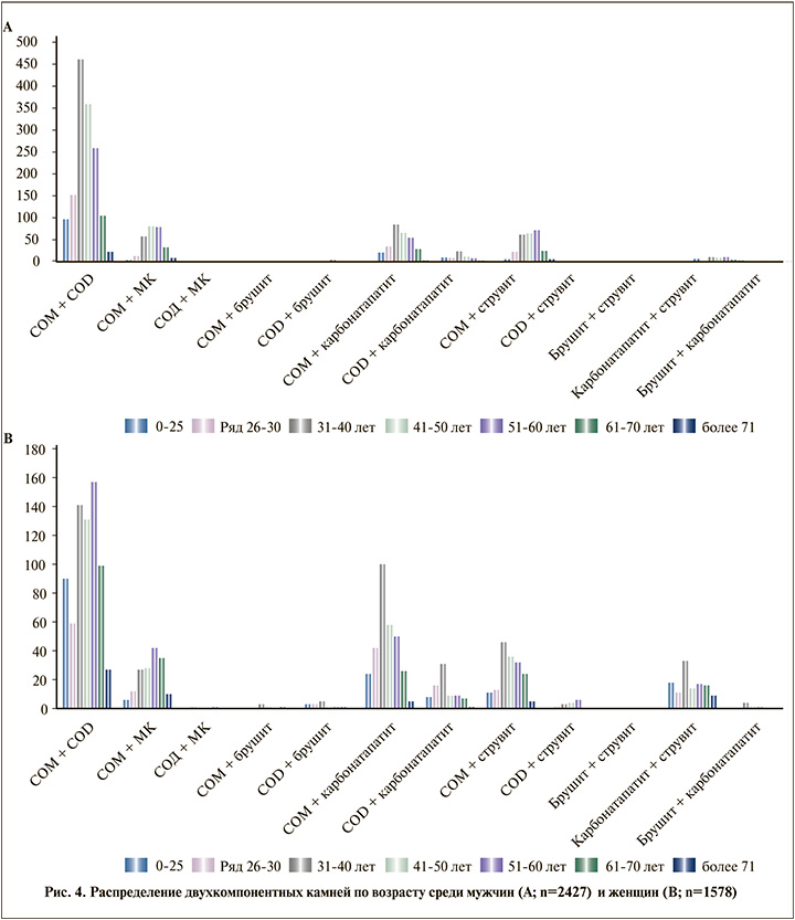
Среди двухкомпонентных камней у взрослых наиболее часто встречались смесь СОМ+СОD – 53,9%, СОМ+СаР – 15%, СОМ+МК – 10,9%, СОМ+струвит – 10,6%. Присоединение инфекционного компонента отмечено в 34,3% всех случаев.
У детей также превалировали камни из СОМ+СОD – 62,8%, СаОх+СаР – 21,4%, СаОх+струвит – 6%, СаР+струвит – 8,4%. Повсеместно наиболее распространенным сочетанием минералов является СОМ+COD – от 67,3% (Сев.-Кавказский ФО) до 45,7% (Дальневосточный ФО). С приблизительно равной частотой встречается сочетание СОМ+СаР и СОМ+струвит. Смесь СОМ+ МК наиболее часто встречается в Республике Казахстан, Центральном, Уральском, Сибирском, Северо-Кавказском, Северо-Западном ФО и Москве.
Доля камней из смеси кальция (СОМ и СОD) в общей массе составила 53,9%, среди мужчин частота выявления данного сочетания достигала 67,4% против 32,6 среди женщин (p<0,001). Камни из СОМ и СаР составили 15% всех двухкомпонентных камней при почти равномерном распределении среди мужчин и женщин – 49,1 и 50,9% соответственно (р=0,778).
Сочетание СОМ+струвит чаще встречалось среди муж-чин – 60,8%/39,2% (р=0,002). СОD+струвит, СаР+струвит – среди женщин (р<0,001), что скорее всего обусловлено более высокими значениями рН мочи, инфекциями мочевыводящих путей (рис. 3).

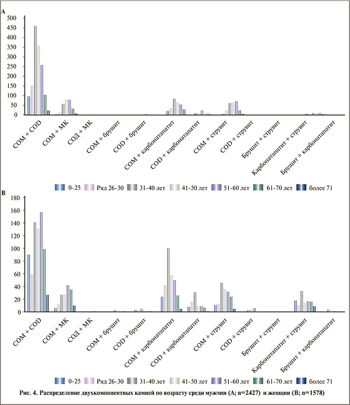
Высокое распространение конкрементов из СОМ и СОD отмечено во всех возрастных группах. Снижение частоты встречаемости данного сочетания отмечается в возрасте старше 70 лет. В возрасте от 26 до 70 лет отмечена высокая встречаемость камней из СОМ и МК (пиком в возрасте 31 – 60 лет). Во всех возрастных группах имеет место высокое распространение смешанных камней из СОМ и СаР, СОМ и струвита, СаР и струвита (присоединение фосфатного компонента) (рис. 4).

Среди детей присоединение фосфатного компонента (брушит, карбонатапатит и струвит) достоверно чаще встречалось у девочек (р<0,001). Сочетание СОМ+СОD немного чаще отмечено у мальчиков (р=0,572).
Среди многокомпонентных мочевых камней (n=2334) самым распространенным было сочетание СОМ+СОD и СаР – 90,4%, следующим по частоте встречаемости в данной группе было сочетание СОМ+СОD и струвита – 6,6%. Сочетание СОМ+СОD и СаР распространено примерно равномерно во всех регионах и составляло от 88,5 до 100% исследованных камней. Сочетание СОМ+СОD и струвита встречается почти равномерно во всех регионах (от 3% (в Южном ФО) до 11,8% (Приволжский ФО). При оценке распределения многокомпонентных мочевых камней среди мужчин и женщин (1408/926) сочетание СОМ+СОD и (СаР) чаще встречалось среди мужчин – 60,2% против 39,8 у женщин (р=0,005). Сочетание СОМ+СОD и струвита преобладало среди мужчин – 60,1% против 39,9 у женщин (р=0,005). Остальные типы многокомпонентных камней встречались с частотой от 0,8 до 1,3% исследований с некоторым преобладанием среди мужчин.
При оценке распределения многокомпонентных камней по возрастным группам отмечено, что смесь СОМ+СОД и СаР встречалась во всех возрастных группах, максимальные показатели среди мужчин в возрасте 30–50 лет, среди женщин в возрасте 30–40 лет, при стабильных показателях в возрасте 40–60 лет. Камни из СОМ+СОД и струвита наиболее часто определяются в возрасте 31–60 лет. По-видимому, формирование данных каменных композиций связано с образованием начального камня на бляшке Рендалла при наличии множественных метаболических нарушений и последующем присоединении воспалительного процесса или ощелачивании мочи по различным причинам, а также бесконтрольного приема антибактериальных препаратов и роста региональной резистентности к ним. Примесь мочевой кислоты к кальций-оксалатному конкременту наиболее часто встречается в возрасте 41–70 лет.
У детей в целом определяется сходная ситуация: наиболее распространенным многокомпонентным по составу камнем являлась смесь СОМ+СОD и СаР – 88,2%, вторыми по частоте встречаемости были камни из смеси СОМ+СОD и струвита – 9,9%. Смесь СОМ+СОD и СаР встречалась почти равномерно среди мальчиков (50,3%) и девочек (49,7%), камни из смеси СОМ+СОD и струвита достоверно чаще встречалась у девочек – 61,9% против 38,1 мальчиков (p<0,001).
Обсуждение. Мочекаменная болезнь распространена во всех регионах планеты. И за последние десятилетия имеет выраженную тенденцию к существенному прогрессированию как в развитых, так и в развивающихся странах. Указанная тенденция обусловлена множественными причинами, включая накопление как генетических факторов развития заболевания, так и факторов, связанных с изменением множественных экологических влияний. Экономические расчеты свидетельствуют о прогрессивном росте затрат на лечение МКБ. По оценкам проекта «Урологические заболевания в Америке», проведенного в 2012 г., стоимость лечения МКБ в настоящее время достигает 10 млрд долл., что делает МКБ одним из самых дорогих урологических заболеваний [1]. Предварительные расчеты предполагают увеличение затрат на лечение МКБ в США к 2030 г. до 15 млрд [2], при этом указанные расчеты не включают финансовых потерь «из-за потери трудоспособности». Исследования, проведенные в Великобритании, показывают, что затраты на лечение МКБ сопоставимы со стоимостью первичного комбинированного лечения рака мочевого пузыря и предстательной железы во всей Великобритании [3].
Многолетние наблюдения указывают на более высокую заболеваемость МКБ среди мужчин. Однако за последние десятилетия отмечается существенное изменение гендерных особенностей заболеваемости МКБ [4]. Имеются свидетельства различий состава мочевых камней в различных возрастных группах [5, 6]. На заболеваемость и распространенность МКБ кроме вышеперечисленных факторов камнеобразования влияют сезонные колебания температуры, генетика, вода, температура окружающей среды, широта, загрязнение, достаток, доступность технологий, пищевые привычки и образ жизни [7–11].
Одним из ведущих факторов принятия решений по метафилактике и лечению МКБ является химический состав мочевого камня. Полученные нами данные исследования показывают, что однокомпонентные мочевые камни встречаются не часто: у 6,6% взрослых и у 5,2% детей. Однако в литературе имеются указания, что однокомпонентные камни встречаются у 30% детей, преимущественно состоят из кальция оксалата [12]. В исследованной нами популяции преобладали смешанные двух- и многокомпонентные мочевые камни. Этот факт безусловно указывает на необходимость инструментального исследования удаленных или отошедших мочевых камней. Клинические рекомендации большинства урологических ассоциаций мира предлагают изучать состав мочевого камня с помощью инфракрасной спектроскопии или рентгеновской дифракции. Эти методы позволяют определять химическую природу составляющих камня и давать полуколичественную оценку их соответствующих пропорций. Визуальное определение состава, особенно многокомпонентных мочевых камней даже с использованием эндоскопических методик недостаточно информативно и требует дальнейшего изучения. Проведенные исследования свидетельствуют, что эндоскопическое распознавание камней представляет собой серьезную проблему. Лишь немногие урологи обладают достаточным опытом, чтобы проводить диагностику, сравнимую с морфо-конституциональным анализом [13, 14].
К сожалению, в настоящее время в случае проведения мелкодисперсной фрагментации камней применение морфо-конституционального метода, предложенного M. Daudon et al. [15], не представляется возможным [15]. Так как этот метод подразумевает комплексный подход, сочетающий тщательное морфологическое исследование поверхности и среза камней с подробным анализом спектроскопических данных местоположения минералов, кристаллической фазы и соответствующей пропорции компонентов камня. Белорусские исследователи предложили метод качественного экспресс-анализа мочевого камня, основанный на комбинированной оптической макроскопии образца, качественных химических реакциях и химических реакциях повторного отложения с последующей микроскопией образовавшегося кристалла [16]. Данная методика показала доступность реализации, малые финансовые затраты и достаточную информативность. В настоящее время появился и развивается новый междисциплинарный подход к изучению мочевых камней – GeoBioMed, объединяющий области геологии, биологии и медицины, который показывает, что камни в почках, состоящие из минералов, богатых кальцием, выпадают в результате совокупности повторяющихся событий кристаллизации, растворения и перекристаллизации, являющихся результатом тех же фундаментальных природных процессов, что и управляли биоминерализацией миллиарды лет на Земле. Идеи GeoBioMed определяют ранее не изученные стратегии разработки и тестирования новых клинических методов лечения для профилактики и лечения камней в почках [17].
Среди исследованных камней преобладали камни, полученные от пациентов мужского пола, и камни с преобладающим содержанием кальция. Определяются особенности типов камнеобразования в различных обследованных регионах, по-видимому обусловленные климатическими и пищевыми особенностями, а также гендерной принадлежностью. Особое значение имеет исследование состава мочевых камней у детей, так как дети с нефролитиазом представляют собой многолетнюю угрозу повторного появления камней в течение жизни (высокий риск рецидива). Частота риска развития рецидива у них достигает 50% через 3 года наблюдения [18, 19].
Информация о химическом составе камня позволяет предположить характерные метаболические нарушения у конкретного пациента. Преобладание моногидратной формы кальция оксалата (COM) связано с повышенным содержанием оксалата в моче, тогда как дигидратная форма (COD) обусловлена гиперкальциурией. Исследование J. E. Brinkman et al. [20] показывает, что однокомпонентный состав конкрементов коррелирует с определенными характеристиками мочи и клиническими данными. Пациенты с СаР, брушитом и дигидратом CaOx моложе и имеют более высокие показатели гиперкальциурии, чем пациенты с моногидратом CaOx и мочевой кислотой. Среди пациентов с камнями из мочевой кислоты преобладают мужчины, тогда как среди пациентов с камнями из СаР больше женщин. У пациентов с брушитом отмечается самая высокая распространенность первичного гиперпаратиреоза [20]. Полученные данные ориентировочно позволяют определять диетические и/или фармакологические рекомендации и помогать в принятии эмпирических клинических решений. В настоящее время известно, что сложное множество факторов, отличных от простой химии мочи, влияет на образование камней, включая продукты деградации белка, клеточные реакции и даже вероятное присутствие микробиома почек [21, 22]. Более того, различные состояния здоровья человека, включая обезвоживание, беременность, диетические пристрастия, диабет, гипертонию, ожирение, генетику и климат, также влияют на образование камней в почках [7, 8, 23–26]. Данные факты являются дополнительным свидетельством необходимости исследования состава мочевых камней и проведения полноценной индивидуальной метаболической оценки с целью выполнения эффективного метафилактического лечения [7, 8, 23–26].
Заключение. Анализ камней имеет хотя и ограниченную, но потенциально значимую роль в упрощенной оценке метаболических нарушений у конкретного пациента. Камни с преобладанием оксалата кальция являются наиболее распространенным типом камней. Данное обстоятельство может свидетельствовать о широком распространении нарушений кальциевого обмена, обусловленных множеством причин. Однако регион проживания, возраст и пол оказывают заметное влияние на тип образующегося камня. Знание общих тенденций изменения типов камнеобразования среди мужчин и женщин в зависимости от возраста может играть важную роль в определении подходов к метафилактике. Изучение распространенности видов мочевых камней на различных территориях страны и мира играет важную роль в предсказании нагрузки на системы здравоохранения в целом и урологическое сообщество в частности, в том числе и в плане расчетов вероятности рецидивирования заболевания и затрат на лечение. От состава мочевых камней зависит выбор адекватной тактики эффективной метафилактики. Увеличение объемов исследования мочевых камней может представить значительную информацию об имеющихся метаболических нарушениях и сопутствующих заболеваниях конкретного пациента.


